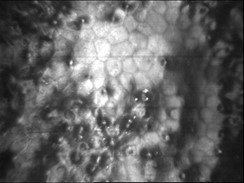
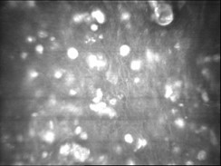
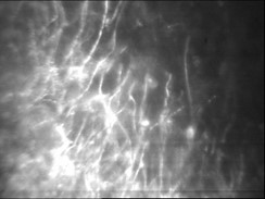
Confocal scan
Confocal microscopy is a modern, innovative, and fast technique to examine the cellular structure of different layers of the cornea, from the most posterior to the most superficial. The images obtained in this way in real time and in vivo from the patient's cornea without the need for tissue sampling are almost similar to the images obtained by light microscopy from tissue sections in vitro.
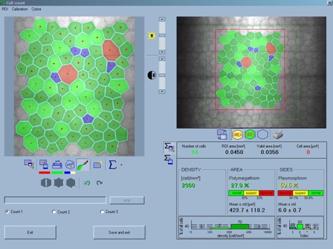
The uses and capabilities of confocal scanning are numerous. Imaging of infectious corneal wounds and examining them for pathogens such as fungus, amoeba and nocardia, diagnosis and evaluation of various corneal dystrophies, especially focal endothelial dystrophy, PPMD and ICE, help in diagnosing the nature of corneal deposits, counting endothelial cells. specular microscopy), counting and determining the distribution of keratocytes, counting the cells of the basal layer of the epithelium, measuring the thickness of the cornea, checking the health status of the subepithelial nerve network and stromal nerves, determining the depth of corneal opacities, evaluating the interface in layer connections Examining the condition of the lenticule in the posterior lamina graft and determining the haziness in corneal opacities are among its capabilities. Confocal scanning can be used as a complement to impression cytology sampling in the diagnosis of corneal squamous cell malignancy.
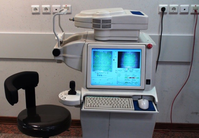
Performing a confocal scan and working with the relevant device requires special skills and expertise, and beyond that is the interpretation of findings and images, which is not possible without acquiring relevant specialized knowledge. Central Eye Bank of the Islamic Republic of Iran is the reference base and the most active confocal scanning center in the country, which has been receiving referred patients from all over the country since 2013. This special service in this center is offered to patients by ophthalmology/eye pathology specialists.